Parable is developed by and for SCI-based clinicians with a variety of features to support complex, long-term cases, such as CARF-oriented documentation, CAPI/HAPI tracking, and powerful 2D and 3D scans with automatic wound and surface area measurement.
Parable's mobile app offers flexible workflows and syncs data to an integrated web dashboard featuring at-a-glance case summaries, report exports, and collaborative monitoring tools.
Learn more about Parable features and capabilities geared for SCI:
- Wound management dashboard with per-wound overviews
- 2D and 3D scanning with automatic metrics, including real surface area
- Detailed and streamlined Pressure Injury documentation with CAPI/HAPI tracking
Wound Management
In Parable, you can document, monitor, and manage each wound belonging to a patient individual dashboard for each wound site belonging to a patient. For complex cases with multiple wounds, the assessment graph provides an at-a-glance overview, with an assessment timeline that lets you drill down to the details of each encounter.
Each wound has its own Gallery, Notes and Communications page, and Audit Log. Individual wound sites can be closed, or re-opened if they require attention again. Reports can be generated at a variety of levels, such as for individual encounters, or for all your wounds in aggregate (such as the the Non-Healing Wound Report). You can quickly visualize a wound's trajectory by reviewing the gallery, which features a timelapse view to showcase progression and results to care team members.
Dashboards provide population-level insights, highlighting the types of cases being seen, recent activity, healing rates, and more.
2D and 3D Scans: Automatic Measurements + Surface Area
Parable features two different scanning mode that produce automatic measurements, including real wound surface area, a key metric for wound healing progression. Wound area measurements provide an objective assessment of wound healing¹, and digital imaging and digital planimetry are the most accurate and reliable methods for measuring wound area²--Parable delivers these capabilities in a highly portable, integrated app with secure cloud sync.
3D Smart Scans use a lightweight (99 grams) 3D sensor attachment for iPad to automatically measure all wound dimensions: length, width, and depth, along with real surface area and volume, generating a topographical, depth-mapped 3D model that you can review in realtime.
Smart Photo 2.0, available on iPhone and iPad, automatically measures length and width in realtime, and generates a real surface area measurement. The Smart Photo process features in-app scanning guidance, and does not require external hardware.
Real surface area measurements more accurately capture the true size of the wound compared to 'manual' length by width estimates. In the example below, from a Parable 3D Smart Scan, the real measured surface area outline in blue (71.9cm²) is significantly lower than the 'manual' length by width surface area would be (10.9 x 10.4 = 113.3cm²).
Accurate surface area measurements provide the best insight into wound progression. Baseline pressure injury size is a significant predictor for healing at 4 weeks³, and wounds with less than 50% surface area reduction in 4 weeks are less likely to heal⁴. Parable's Non-Healing Wound Report provides insight into wound progression based on surface area.
CAPI/HAPI Tracking and CARF Pressure Injury Documentation
For advanced monitoring of pressure injuries, you can indicate the staging and origin when adding a pressure injury wound site in Parable. Stage and origin can be updated at any time by editing the wound site.
To help streamline documentation of CARF accreditation requirements, the pressure injury assessment in Parable includes a fully digitized SCI-PUMT protocol with additional questions to address CARF factors, including surrounding skin details, barriers to healing, discharge planning, and more.
Digitized clinical assessments protocols like SCI-PUMT or Braden Scale produce qualitative assessment scores which are plotted along with wound metrics on the overview graph, can provide research-backed insight into wound progression. Parable streamlines documentation and scoring of clinical assessment protocols down to a few taps.
Facet Tracking and Location Tracking
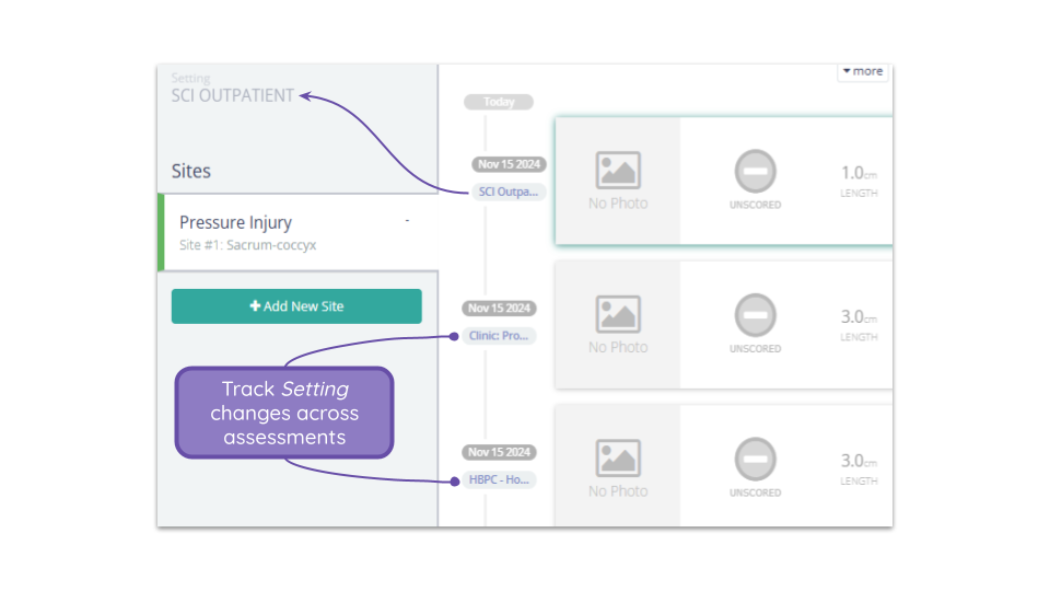
For long-term SCI patients that may receive care in a variety of settings, Parable makes it easy to follow where a patient was seen in addition to when.
If your team uses multiple Settings in Parable (for example, if you have separate sub-Regions in Parable for SCI Inpatient, SCI Outpatient, and SCI Home Care), you can update the patient's Setting during each encounter. On the Assessment Timeline, you'll see a label indicating the Setting in which the encounter took place.
For teams with different Setting configurations, or no Settings, Location Tracking allows you to label any encounter as Inpatient, Outpatient, or Home Health. These location labels will show up on the Patient List, Patient Profile, and Assessment Timeline.
CPRS/VistA Integration
You can import your EHR patients directly into Parable by using familiar search patterns or by scanning a patient ID barcode.
Automatic note creation (e-filed transfers) of Parable images and assessment data to the EHR can also be enabled. For more information, please see CPRS/VistA: Quick Guide [Non-Consult].
Modality
Parable is a mobile app available on iPhone or iPad, and can be used on existing, multi-purpose devices. A standalone device is not required, and all imaging and documentation processes take place in one single Parable app.
The 3D sensor attachment required for 3D Smart Scans weighs only 99 grams, making it highly portable in any context.
The 3D sensor is only required for 3D scans--all other platform and app features (including 2D Smart Photos), do not require a sensor. Note that the 3D sensor works exclusively with iPads and is not compatible with iPhones.
There are no patient or wound site limits, and as many Supporting Photos as needed can be added each check-in.
References
- Darwin, Evan S et al. “Comparison of 3-dimensional Wound Measurement With Laser-assisted and Hand Measurements: A Retrospective Chart Review.” Wound management & prevention vol. 65,1 (2019): 36-41.
- Jørgensen, Line Bisgaard et al. “Methods to assess area and volume of wounds - a systematic review.” International wound journal vol. 13,4 (2016): 540-53. doi:10.1111/iwj.12472
- Guihan, Marylou et al. “Difficulty in Identifying Factors Responsible for Pressure Ulcer Healing in Veterans With Spinal Cord Injury.” Archives of physical medicine and rehabilitation vol. 97,12 (2016): 2085-2094.e1. doi:10.1016/j.apmr.2016.05.025
- Coerper, Stephan et al. “Fifty percent area reduction after 4 weeks of treatment is a reliable indicator for healing--analysis of a single-center cohort of 704 diabetic patients.” Journal of diabetes and its complications vol. 23,1 (2009): 49-53. doi:10.1016/j.jdiacomp.2008.02.001